High-Res Microscope May Make Cervical Cancer Detection Easier

There’s a new technology in the works that may, one day, make screening for cervical cancer faster, easier, and more precise. The tool is a high-resolution microscope that makes it possible to visualize and analyze individual cells instantaneously using very fast pulses of light.
The researchers who are working on it hope to overcome some of the limitations of the Pap smear, with the goal of fewer patients having to endure biopsies, additional testing, or the stress of waiting for a specialist to analyze the results.
Every year, millions of women undergo a Pap smear to look for early cervical cancers or pre-cancerous changes in the cervix. The test was introduced in the late 1940s and has helped to significantly reduce mortality from cervical cancer in the decades since. In the U.S. alone, the death rate from cervical cancer has dropped more than 50% in the last 30 years thanks mostly to increased use of Pap tests.
This test, however, has limitations. For example, a specialist must use his or her judgment to visually determine if cells look abnormal, which can lead to false positives. Because of this, women with an abnormal Pap result must often get another Pap or an HPV test to check for a human papilloma virus infection – the cause of most cervical cancers. Even then, some women need a biopsy (removal of tissue), to determine if in fact a pre-cancer or cancer exists.
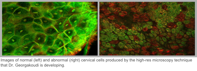
This “can cause a lot of unneeded cost and stress,” says Irene Georgakoudi, Ph.D., a biophysicist at Tufts University, who is working with Karl Munger, Ph.D., a Tufts University virologist, on a more reliable and less invasive way of detecting cancers, such as cervical, early.
Early Signs of Cancer in Cells Often Too Small for Existing Technology to Pick Up On
Georgakoudi hopes her optical technique will eventually reduce the need for biopsies. Georgakoudi’s new tool, which she is developing with the help of funding from the American Cancer Society, would allow for an automated analysis of results, removing “human subjectivity from the interpretation process.”
Georgakoudi says the very early changes that occur to cells before cancer invasion or metastasis can be “truly microscopic.” So small that even when doctors use existing magnification technology they may not be able to see the types of cell changes that would alert them to the development of cancer.
“On the other hand, there are other things on the surface of the cervix besides pre-cancerous changes that make many tissue areas look suspicious, so the doctor will biopsy those areas and many times it turns out not to be cancer,” says Georgakoudi.
She explains that the situation is similar for other types of cancer, such as those in the oral cavity and colon. Sometimes, preliminary screening tests may miss signs of cancer or pre-cancer if they are too small – and other times what a doctor thinks is an abnormality may end up to not be, leading to “too many biopsies of normal tissue; costing a lot of money and putting patients through a lot of agony,” says Georgakoudi.
“The goal for the researchers in my field is to develop techniques that basically allow us to identify microscopic changes at an early stage that are both more sensitive and more specific than current tools allow, so doctors won’t need to biopsy tissue if there is a good chance the tissue is not going to be abnormal.”
A New Super-Sensitive Microscope May Be Able to See What Current Tools Cannot
Georgakoudi is working on a high-resolution microscope whose magnification power far outstrips that of a colposcope, the tool currently used to examine the cervix to look for cancer. “Instead of visualizing the surface of the cervix at 6 to 15 times magnification we are looking at magnifications that are on the order of 25 to 60 times, so we can really visualize individual cells.”

It also allows users to very specifically select the layer of tissue they want to view. Georgakoudi says this is possible through the use of a certain kind of light, which enables her to identify if cells have undergone the type of changes indicative of cancer.
“In cells we have two enzymes that if you shine light of the right color on them they absorb the light and then reemit light of a slightly different color.” These two enzymes are important because they are involved in cellular processes that change when cancer is developing. So by measuring the light the enzymes reemit, Georgakoudi says her new tool will be able to assess whether cancerous changes are occurring.
Guiding Biopsy and Beyond
“The first goal is to use this technology as a guide to biopsy,” says Georgakoudi. She means that doctors would use the high-powered microscope to help them see which parts of tissue to remove so that they don’t miss the abnormal areas.
If she is able to show that her tool works well for this, Georgakoudi believes doctors could use it prior to biopsy to determine whether there are in fact any suspicious areas that truly need to be removed for further examination. Because Georgakoudi’s optical technique is more specific and sensitive than a colposcope, she hopes it will reduce the need for biopsies, which can be painful and stressful.
“The Holy Grail is to not have to go for the biopsy or at least in some cases replace biopsy.”
Thinking even bigger, Georgakoudi says there may even be uses for the technology during breast or brain cancer surgery. “In principle, you could use a high-res technology like this to image the margins of the tissue you removed to make sure all cancer has been removed, which is very important because even if you leave a few cells behind a tumor can grow.”
Georgakoudi says her testing in real human tissue samples is thus far promising. She is now working on building a larger database of human tissues for further testing to help refine and perfect the technology.
American Cancer Society news stories are copyrighted material and are not intended to be used as press releases. For reprint requests, please see our Content Usage Policy.