Your gift is 100% tax deductible
What Is Testicular Cancer?
Cancer that starts in the testicles is called testicular cancer. Cancer starts when normal cells begin to grow out of control. To understand testicular cancer, it helps to know about the normal structure and function of the testicles.
What are testicles?
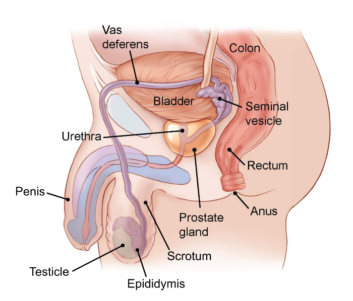
Testicles (also called testes; a single testicle is called a testis) are part of the male reproductive system. The 2 organs are each normally a little smaller than a golf ball in adult males. They are held within a sac of skin called the scrotum. The scrotum hangs under the base of the penis.
Testicles have 2 main functions:
- They make male hormones (androgens) like testosterone.
- They make sperm, the male cells needed to fertilize a female egg cell to start a pregnancy.
Sperm cells are made in long, thread-like tubes inside the testicles called seminiferous tubules. They are then stored in a small, coiled tube behind each testicle called the epididymis. This is where they mature.
During ejaculation, sperm cells are carried from the epididymis through the vas deferens to the seminal vesicles. There, they mix with fluids made by the vesicles, prostate gland, and other glands to form semen. This fluid then enters the urethra, the tube in the center of the penis through which both urine and semen leave the body.
Types of testicular cancer
The testicles are made up of many types of cells, each of which can develop into one or more types of cancer. It's important to know the type of cell the cancer started in and what kind of cancer it is because they differ in how they're treated and in their prognosis (outlook).
Doctors can tell what type of testicular cancer you have by looking at the cells under a microscope.
Germ cell tumors
More than 90% of cancers of the testicle start in cells known as germ cells. These are the cells that make sperm. The main types of germ cell tumors (GCTs) in the testicles are seminomas and non-seminomas.
These types occur about equally. Many testicular cancers contain both seminoma and non-seminoma cells. These mixed germ cell tumors are treated as non-seminomas because they grow and spread like non-seminomas.
Seminomas tend to grow and spread more slowly than non-seminomas. The 2 main sub-types of these tumors are classical (or typical) seminomas and spermatocytic seminomas.
- Classical seminoma: More than 95% of seminomas are classical. These usually occur in men between 25 and 45.
- Spermatocytic seminoma: This rare type of seminoma tends to occur in older men. The average age is about 65. Spermatocytic tumors tend to grow more slowly and are less likely to spread to other parts of the body than classical seminomas.
Some seminomas can increase blood levels of a protein called human chorionic gonadotropin (hCG). hCG can be checked with a simple blood test and is considered a tumor marker for certain types of testicular cancer. It can be used for diagnosis and to check how the patient is responding to treatment.
These types of germ cell tumors usually occur in men between their late teens and early 30s. The 4 main types of non-seminoma tumors are embryonal carcinoma, yolk sac carcinoma, choriocarcinoma, and teratoma. Most tumors are a mix of different types (sometimes with seminoma cells too), but this doesn’t change the treatment of most non-seminoma cancers.
Embryonal carcinoma: These cells are found in about 40% of testicular tumors, but pure embryonal carcinomas occur only 3-4% of the time. When seen under a microscope, these tumors can look like tissues of very early embryos. This type of non-seminoma tends to grow rapidly and spread outside the testicle.
Embryonal carcinoma can increase blood levels of a tumor marker protein called alpha-fetoprotein (AFP), as well as human chorionic gonadotropin (hCG).
Yolk sac carcinoma: These tumors are so named because their cells look like the yolk sac of an early human embryo. Other names for this cancer include yolk sac tumor, endodermal sinus tumor, infantile embryonal carcinoma, and orchidoblastoma.
This is the most common form of testicular cancer in children (especially in infants), but pure yolk sac carcinomas (tumors that do not have other types of non-seminoma cells in them) are rare in adults. When they occur in children, these tumors usually are treated successfully. But they're of more concern when they occur in adults, especially if they are pure. Yolk sac carcinomas respond very well to chemotherapy, even if they have spread.
This type of tumor almost always increases blood levels of AFP.
Choriocarcinoma: This is a very rare and fast-growing type of testicular cancer in adults. Pure choriocarcinoma is likely to spread rapidly to other parts of the body, including the lungs, bones, and brain. More often, choriocarcinoma cells are seen with other types of non-seminoma cells in a mixed germ cell tumor. These mixed tumors tend to have a somewhat better outlook than pure choriocarcinomas, although the presence of choriocarcinoma is always a worrisome finding.
This type of tumor increases blood levels of hCG.
Teratoma: Teratomas are germ cell tumors with areas that, under a microscope, look like each of the 3 layers of a developing embryo: the endoderm (innermost layer), mesoderm (middle layer), and ectoderm (outer layer). Pure teratomas of the testicles are rare and do not increase AFP or hCG levels. Most often, teratomas are seen as parts of mixed germ cell tumors.
There are 3 main types of teratomas:
- Mature teratomas are tumors formed by cells that are similar to the cells of adult tissues. They rarely spread. They can usually be cured with surgery, but some come back (recur) after treatment.
- Immature teratomas are less well-developed cancers with cells that look like those of an early embryo. This type is more likely than a mature teratoma to grow into (invade) nearby tissues, spread (metastasize) outside the testicle, and come back (recur) years after treatment.
- Teratomas with somatic type malignancy are very rare. These cancers have some areas that look like mature teratomas but have other areas where the cells have become a type of cancer that normally develops outside the testicle (such as a sarcoma, adenocarcinoma, or even leukemia).
Carcinoma in situ of the testicle
Testicular germ cell cancers can start as a non-invasive form of the disease called carcinoma in situ (CIS) or intratubular germ cell neoplasia. In testicular CIS, the cells look abnormal under the microscope, but they have not yet spread outside the walls of the seminiferous tubules (where sperm cells are formed). Carcinoma in situ doesn’t always progress to invasive cancer.
It's hard to find CIS before it becomes an invasive cancer because it generally doesn't cause symptoms or form a lump that you or the doctor can feel. The only way to diagnose testicular CIS is to have a biopsy. (This is a procedure to take out a tiny bit of tissue so it can be checked under a microscope.) Sometimes CIS is found incidentally (by accident) when a testicular biopsy is done for another reason, such as infertility.
Experts don’t agree about the best treatment for CIS. Since CIS doesn’t always become an invasive cancer, many doctors in the United States consider observation (watchful waiting) to be the best treatment option.
When CIS of the testicle becomes invasive, its cells are no longer just in the seminiferous tubules, they've grown into other structures of the testicle. These cancer cells can then spread either to the lymph nodes (small, bean-shaped collections of white blood cells) through lymphatic vessels (tiny fluid-filled tubes that connect the lymph nodes), or through the blood to other parts of the body.
Stromal tumors
Tumors can also start in the supportive and hormone-producing tissues, or stroma, of the testicles. These tumors are known as gonadal stromal tumors. They make up less than 5% of adult testicular tumors, but up to 20% of childhood testicular tumors. The main types are Leydig cell tumors and Sertoli cell tumors.
These tumors start in the Leydig cells in the testicle that normally make male sex hormones (androgens like testosterone). Leydig cell tumors can develop in both adults and children. These tumors often make androgens (male hormones), but sometimes they make estrogens (female sex hormones).
Most Leydig cell tumors are not cancer (benign). They seldom spread beyond the testicle and can often be cured with surgery. Still, a small number of Leydig cell tumors do spread to other parts of the body. These tend to have a poor outlook because they usually don't respond well to chemo or radiation therapy.
These tumors start in normal Sertoli cells, which support and nourish the sperm-making germ cells. Like the Leydig cell tumors, these tumors are usually benign. But if they spread, they usually don’t respond well to chemo or radiation therapy.
Secondary testicular cancers
Cancers that start in another organ and then spread (metastasize) to the testicle are called secondary testicular cancers. These are not true testicular cancers – they don't start in the testicles. They're named and treated based on where they started.
Lymphoma is the most common secondary testicular cancer. Testicular lymphoma is more common in men older than 50 than primary testicular tumors. The outlook depends on the type and stage of lymphoma. The usual treatment is surgical removal, followed by radiation and/or chemotherapy.
In boys with acute leukemia, the leukemia cells can sometimes form a tumor in the testicle. Along with chemotherapy to treat the leukemia, this might require treatment with radiation or surgery to remove the testicle.
Cancers of the prostate, lung, skin (melanoma), kidney, and other organs also can spread to the testicles. The prognosis for these cancers tends to be poor because these cancers have usually spread widely to other organs as well. Treatment depends on the specific type of cancer.
- Written by
- References

Developed by the American Cancer Society medical and editorial content team with medical review and contribution by the American Society of Clinical Oncology (ASCO).
National Cancer Institute. Testicular Cancer Treatment (PDQ®)–Patient Version. May 17, 2023. Accessed at www.cancer.gov/types/testicular/patient/testicular-treatment-pdq on June 1, 2025.
National Comprehensive Cancer network. NCCN Clinical Guidelines in Oncology (NCCN Guidelines). Testicular Cancer. Version 1.2025 – Jan 17, 2025. Accessed at https://www.nccn.org on Feb 18, 2025.
Last Revised: August 10, 2025
American Cancer Society medical information is copyrighted material. For reprint requests, please see our Content Usage Policy.
American Cancer Society Emails
Sign up to stay up-to-date with news, valuable information, and ways to get involved with the American Cancer Society.