Your gift is 100% tax deductible
Gastrointestinal Neuroendocrine Tumor Stages
After someone is diagnosed with a gastrointestinal (GI) neuroendocrine tumor, doctors will try to figure out if it has spread, and if so, how far. This process is called staging. The stage of cancer describes how much cancer is in the body. It helps determine how serious the cancer is and how best to treat it. Doctors also use a cancer's stage when talking about survival statistics.
How is the stage determined?
GI neuroendocrine tumors are typically given a clinical stage based on the results of any exams, biopsies, and imaging tests (as described in Tests for Gastrointestinal Neuroendocrine Tumors. If surgery has been done, the pathologic stage (also called the surgical stage) can also be determined.
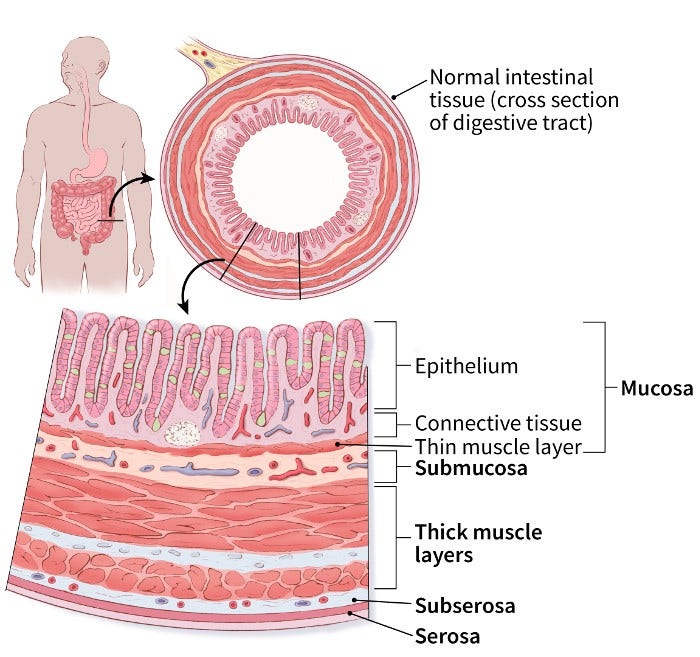
GI neuroendocrine tumors typically start in the inner lining of the wall of the GI tract. As they grow, they can spread into deeper layers of the GI tract. For most of the GI tract, these layers include:
- Mucosa: This is the innermost layer. It has 3 parts: the top layer of cells (the epithelium), a thin layer of connective tissue (the lamina propria), and a thin layer of muscle (the muscularis mucosa).
- Submucosa: This is the fibrous tissue that lies beneath the mucosa.
- Thick muscle layer (muscularis propria): This layer of muscle contracts to force the food along the GI tract.
- Subserosa and serosa: These are the thin outermost layers of connective tissue that cover the GI tract. The serosa is also known as the visceral peritoneum.

Localized, regional, and distant stages
Until recently, there was no standard staging system for describing the spread of GI neuroendocrine tumors. Many doctors simply staged GI neuroendocrine tumors as localized, regional spread, and distant spread. This approach was fairly easy to understand and could be useful when determining treatment options.
- Localized: The cancer has not spread beyond the wall of the organ it started in (for example, the stomach, small intestine, or rectum).
- Regional spread: The cancer has either spread to nearby lymph nodes, or it has grown through the wall of the organ where it started and into nearby tissues such as fat, ligaments, and muscle (or both).
- Distant spread: The cancer has spread to tissues or organs that are not near where the cancer started (such as the liver, bones, or lungs).
The AJCC TNM staging system
The staging system most often used for GI neuroendocrine tumors is the American Joint Committee on Cancer (AJCC) TNM system, which is based on 3 key pieces of information:
- The size and extent of the main tumor (T): Where is the tumor? How far has it grown into the wall of the GI tract and nearby structures?
- The spread to nearby lymph nodes (N): Has the cancer spread to nearby lymph nodes?
- The spread (metastasis; M) : Has the cancer spread to distant parts of the body? The most common sites of spread are lymph nodes far away from the tumor, the liver, the lungs, and the bones.
Numbers or letters after T, N, and M give more details about each of these factors. Higher numbers mean the cancer is more advanced.
Once the T, N, and M categories of the cancer have been determined, this information is combined in a process called stage grouping to assign an overall stage. For more information, see Cancer Staging.
The main stages of GI neuroendocrine tumors in the TNM system range from I (1) through IV (4). Some stages might be divided further with letters (A, B, etc.). As a rule, the lower the number, the less the cancer has spread. A higher number, such as stage IV, means cancer has spread more. And within a stage, an earlier letter means a lower stage. Although each person’s cancer experience is unique, cancers with similar stages tend to have a similar outlook and are often treated in much the same way.
The system described below includes lower-grade neuroendocrine tumors that start in the GI tract, but not other types of cancers that can start there. (For example, it doesn't include high-grade neuroendocrine carcinomas, or the more common types of stomach cancer or colorectal cancer, which have their own staging systems.)
The stages of GI neuroendocrine tumors are slightly different, based on which part of the digestive tract the cancer starts in:
- The stomach
- The small intestine (jejunum or ileum)*
- The appendix
- The colon or rectum
*Neuroendocrine tumors starting in the duodenum or ampulla of Vater are uncommon and have their own staging system, which is not included here.
GI neuroendocrine tumor staging with the TNM system can be complex. If you have any questions about your cancer's stage or what it means, ask your doctor to explain it to you in a way you understand.
Stages of neuroendocrine tumors of the stomach
AJCC stage |
Stage grouping |
Stage description* |
I |
T1 |
The tumor is no more than 1 centimeter (cm) across and has grown from the top layer of cells and into deeper layers, such as the lamina propria or the submucosa (T1). The cancer has not spread to nearby lymph nodes (N0) or to distant parts of the body (M0). |
II |
T2 |
The tumor has grown into the lamina propria or submucosa (or both) and is greater than 1 cm across; OR the tumor has grown into the main muscle layer of the stomach (the muscularis propria) (T2). The cancer has not spread to nearby lymph nodes (N0) or to distant parts of the body (M0). |
OR |
||
T3 |
The tumor has grown through the muscularis propria and into the subserosa (T3). The cancer has not spread to nearby lymph nodes (N0) or to distant parts of the body (M0). |
|
III |
T4 |
The tumor has grown into the outer layer of tissue covering the stomach (the serosa or visceral peritoneum) or into nearby organs or structures (T4). The cancer has not spread to nearby lymph nodes (N0) or to distant parts of the body (M0). |
OR |
||
Any T |
The tumor can be any size and might or might not have grown into nearby structures (any T). It has spread to nearby lymph nodes (N1), but not to distant parts of the body (M0). |
|
IV |
Any T |
The tumor can be any size and might or might not have grown into nearby structures (any T). It might or might not have spread to nearby lymph nodes (any N). The cancer has spread to distant parts of the body (M1). |
Stages of neuroendocrine tumors of the small intestine
(jejunum or ileum)
AJCC stage |
Stage grouping |
Stage description* |
I |
T1 |
The tumor is no more than 1 centimeter (cm) across and has grown from the top layer of cells and into deeper layers, such as the lamina propria or the submucosa (T1). The cancer has not spread to nearby lymph nodes (N0) or to distant parts of the body (M0). |
II |
T2 |
The tumor has grown into the lamina propria or submucosa (or both) and is greater than 1 cm across; OR the tumor has grown into the main muscle layer of the intestine (the muscularis propria) (T2). The cancer has not spread to nearby lymph nodes (N0) or to distant parts of the body (M0). |
OR |
||
T3 |
The tumor has grown through the muscularis propria and into the subserosa (T3). The cancer has not spread to nearby lymph nodes (N0) or to distant parts of the body (M0). |
|
III |
T4 |
The tumor has grown into the outer layer of tissue covering the intestine (the serosa or visceral peritoneum) or into nearby organs or structures (T4). The cancer has not spread to nearby lymph nodes (N0) or to distant parts of the body (M0). |
OR |
||
Any T |
The tumor can be any size and might or might not have grown into nearby structures (any T). It has spread to nearby lymph nodes (N1 or N2), but not to distant parts of the body (M0). |
|
IV |
Any T |
The tumor can be any size and might or might not have grown into nearby structures (any T). It might or might not have spread to nearby lymph nodes (any N). The cancer has spread to distant parts of the body (M1). |
Stages of neuroendocrine tumors of the appendix
AJCC stage |
Stage grouping |
Stage description* |
I |
T1 |
The tumor is no more than 2 centimeters (cm) across (T1). The cancer has not spread to nearby lymph nodes (N0) or to distant parts of the body (M0). |
II |
T2 |
The tumor is more than 2 cm but no more than 4 cm across (T2). The cancer has not spread to nearby lymph nodes (N0) or to distant parts of the body (M0). |
OR |
||
T3 |
The tumor is more than 4 cm across, OR it has grown into the subserosa or the mesoappendix (T3). The cancer has not spread to nearby lymph nodes (N0) or to distant parts of the body (M0). |
|
III |
T4 |
The tumor has grown into the outer layer of tissue covering the appendix (the peritoneum) or into nearby organs or structures (T4). The cancer has not spread to nearby lymph nodes (N0) or to distant parts of the body (M0). |
OR |
||
Any T |
The tumor can be any size and might or might not have grown into nearby structures (any T). It has spread to nearby lymph nodes (N1), but not to distant parts of the body (M0). |
|
IV |
Any T |
The tumor can be any size and might or might not have grown into nearby structures (any T). It might or might not have spread to nearby lymph nodes (any N). The cancer has spread to distant parts of the body (M1). |
Stages of neuroendocrine tumors of the colon or rectum
AJCC stage |
Stage grouping |
Stage description* |
I |
T1 |
The tumor is no more than 2 centimeters (cm) across and has grown from the top layer of cells and into deeper layers, such as the lamina propria or the submucosa (T1). The cancer has not spread to nearby lymph nodes (N0) or to distant parts of the body (M0). |
IIA |
T2 |
The tumor has grown into the lamina propria or submucosa (or both) and is greater than 2 cm across; OR the tumor has grown into the main muscle layer (the muscularis propria) (T2). The cancer has not spread to nearby lymph nodes (N0) or to distant parts of the body (M0). |
IIB |
T3 |
The tumor has grown through the muscularis propria and into the subserosa (T3). The cancer has not spread to nearby lymph nodes (N0) or to distant parts of the body (M0). |
IIIA |
T4 |
The tumor has grown into the outer layer of tissue covering the intestine (the serosa or visceral peritoneum) or into nearby organs or structures (T4). The cancer has not spread to nearby lymph nodes (N0) or to distant parts of the body (M0). |
IIIB |
Any T |
The tumor can be any size and might or might not have grown into nearby structures (any T). It has spread to nearby lymph nodes (N1), but not to distant parts of the body (M0). |
IV |
Any T |
The tumor can be any size and might or might not have grown into nearby structures (any T). It might or might not have spread to nearby lymph nodes (any N). The cancer has spread to distant parts of the body (M1). |
*The following additional categories are not listed in the tables above:
-
TX: Main tumor cannot be assessed due to lack of information.
-
T0: No evidence of a main tumor.
-
NX: Nearby lymph nodes cannot be assessed due to lack of information.
- Written by
- References

Developed by the American Cancer Society medical and editorial content team with medical review and contribution by the American Society of Clinical Oncology (ASCO).
American Joint Committee on Cancer. Neuroendocrine Tumors of the Appendix. In: AJCC Cancer Staging Manual. 8th ed. New York, NY: Springer; 2018: 389-394.
American Joint Committee on Cancer. Neuroendocrine Tumors of the Colon and Rectum. In: AJCC Cancer Staging Manual. 8th ed. New York, NY: Springer; 2017: 395-406.
American Joint Committee on Cancer. Neuroendocrine Tumors of the Jejunum and Ileum. In: AJCC Cancer Staging Manual. 8th ed. New York, NY: Springer; 2017: 375-487.
American Joint Committee on Cancer. Neuroendocrine Tumors of the Stomach. In: AJCC Cancer Staging Manual. 8th ed. New York, NY: Springer; 2017: 351-359.
Last Revised: August 8, 2025
American Cancer Society medical information is copyrighted material. For reprint requests, please see our Content Usage Policy.
American Cancer Society Emails
Sign up to stay up-to-date with news, valuable information, and ways to get involved with the American Cancer Society.


