Your gift is 100% tax deductible.
Brain Tumors in Adults
If you or someone you love has a brain tumor, knowing what to expect can help. Here you’ll find information on brain tumors in adults, including risk factors, symptoms, and how they are found and treated. You’ll also find information about spinal cord tumors, which are similar to brain tumors, but less common. (For information on children’s tumors, see Brain Tumors in Children.)
About adult brain tumors and spinal cord tumors
Brain tumors and spinal cord tumors are masses of abnormal cells in the brain or spinal cord that have grown out of control.
Are brain tumors always cancer?
Brain tumors and spinal cord tumors can be benign (non-cancerous) or malignant (cancer):
- Benign tumors can grow bigger and press on nearby areas. But they don’t grow directly into nearby tissues or spread to other parts of the body, so benign tumors in most parts of the body are almost never life-threatening.
- Malignant tumors (cancers) can grow into (invade) nearby structures and might even spread to other parts of the body.
In most other parts of the body, it is very important to distinguish between benign and malignant tumors. But in the brain, both benign and malignant tumors can cause serious problems and be dangerous, so the difference between them is less important. For this reason, doctors often talk about “brain tumors” instead of “brain cancer.”
How serious are brain tumors and spinal cord tumors?
Brain tumors rarely spread to other parts of the body, but they can still be very serious. As they grow, they can press on or damage nearby parts of the brain. Even “benign” tumors can cause harm as they get bigger, because they can take up space and damage other parts of the brain. This can sometimes be life-threatening.
The main concerns with brain tumors and spinal cord tumors are:
- How fast they grow
- How readily they spread through the rest of the brain or spinal cord
- Where they are located
- Whether they can be removed (or destroyed) and not come back
But both benign and malignant brain and spinal cord tumors can be life-threatening.
Adult vs. childhood brain tumors
Brain and spinal cord tumors tend to be different in adults and children. They often form in different areas, develop from different cell types, and may have a different prognosis (outlook) and treatments.
For more on the different types of adult brain tumors, see Types of Brain Tumors in Adults.
To learn more about tumors in children, see Brain Tumors in Children.
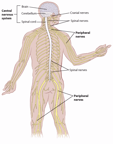
The central nervous system
To understand brain and spinal cord tumors, it helps to know about the normal structure and function of the central nervous system (CNS), which includes the brain and spinal cord. Together, they control how we think, feel, move, and sense the world around us.
- The brain is in charge of thought, memory, emotions, speech, vision, hearing, breathing, movement, and much more.
- The spinal cord connects the brain to the rest of the body. Along with special head nerves called cranial nerves, it carries messages back and forth. These messages tell muscles how to move, bring in signals from our senses, and help control our organs.
Both the brain and spinal cord are well protected, the brain by the skull and the spinal cord by the bones of the spine (vertebrae).
They are also cushioned by a clear liquid called cerebrospinal fluid (CSF). This fluid is made in small structures called the choroid plexus, which sit inside spaces in the brain called ventricles. The ventricles and the spaces around the brain and spinal cord are filled with CSF.
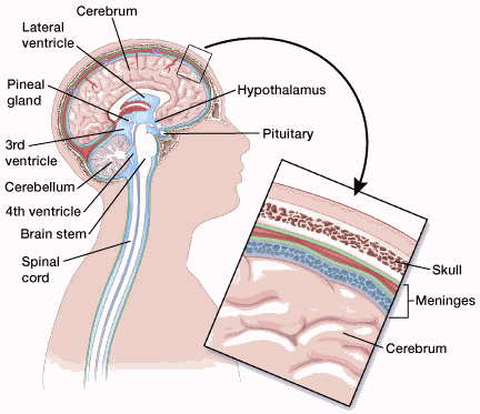
Main parts of the brain
The brain has 3 main areas, each with a special job:
- Cerebrum: The large, upper part of the brain, which is split into left and right halves. It controls thinking, memory, emotions, language, and voluntary movements (like walking, chewing, or throwing a ball). It also processes information from senses like sight, sound, smell, touch, and pain.
- Cerebellum: Located at the back of the brain, below the cerebrum. It helps coordinate balance and movement.
- Brain stem: The lower part of the brain that connects to the spinal cord. It controls basic life functions like breathing and heartbeat. It also carries signals between the brain and the rest of the body. The brain stem has 3 parts: the midbrain, pons, and medulla. Because the brain stem is small but vital, surgery in this area can be very risky.
Cranial nerves
The cranial nerves extend directly from the brain (as opposed to coming out of the spinal cord). They control movement and sensation in the face, eyes, ears, tongue, and some other areas.
Spinal cord
The spinal cord is a long bundle of nerves inside the spine. It carries signals between the brain and the rest of the body related to movement, sensation, and functions like bladder and bowel control.
Pituitary gland and hypothalamus
The pituitary is a small gland at the base of the brain. It is connected to a part of the brain called the hypothalamus. Together, they affect other glands in the body, which in turn influence things like growth, metabolism, sexual development, milk production, and water balance. Tumors or treatments in this area can cause hormone changes. If this happens, people may need to take medicine to replace the missing hormones.
For more on tumors that start in the pituitary, see Pituitary Tumors.
Pineal gland
The pineal gland is a small gland deep inside the brain. It’s part of the body’s hormone system and helps control sleep by making melatonin. Tumors here are rare, but one type is called a pineoblastoma.
Blood-brain barrier
This is a protective layer around the brain’s blood vessels. It helps keep toxins and some other substances in the blood from getting into the brain. Unfortunately, it can also stop many chemotherapy drugs from reaching brain tumors.
Choroid plexus
This is the area of the brain within the ventricles that makes cerebrospinal fluid (CSF), which cushions and nourishes the brain.
Cells and tissues in the brain and spinal cord
The brain and spinal cord are made up of many kinds of cells and tissues. These can develop into different types of tumors.
Neurons (nerve cells): These are the “working” cells in the brain. They control thought, memory, emotion, speech, muscle movement, and sensation, and many other body functions.
Unlike most other types of cells in the body, most neurons in the brain and spinal cord stop dividing about a year after birth, so they rarely form tumors. But neurons can be damaged by tumors that start near them.
Glial cells: Glial cells are the “helper” cells of the brain. Most brain and spinal cord tumors start in glial cells. As a group, these tumors are called gliomas.
There are 3 main types of glial cells:
- Astrocytes support and nourish neurons. They also form scar tissue when the brain is injured. Most tumors starting in these cells are called astrocytomas or glioblastomas.
- Oligodendrocytes make myelin, a fatty coating that insulates nerve fibers and helps signals travel through neurons. Tumors starting in these cells are called oligodendrogliomas.
- Ependymal cells line the ventricles (fluid-filled areas) within the brain and form part of the pathway through which CSF flows. Tumors starting in these cells are called ependymomas.
A fourth type of cell, called microglia, are the infection-fighting cells of the central nervous system. They are part of the immune system, so they’re not truly glial cells, and they rarely form tumors.
Neuroectodermal cells: Also known as primitive neuroepithelial cells, these are early “starter” cells that help form the nervous system during development. They are found throughout the brain, although they are not often seen in the adult central nervous system. The most common tumors that come from these cells develop in the cerebellum and are called medulloblastomas.
Meninges: These are thin layers of tissue that cover and protect the brain and spinal cord. Tumors that start in these tissues are called meningiomas. In adults, they’re among the most common tumors that affect the brain, even though they’re not technically brain tumors.
Quick Guides
- Written by
- References

Developed by the American Cancer Society medical and editorial content team with medical review and contribution by the American Society of Clinical Oncology (ASCO).
Dorsey JF, Salinas RD, Dang M, et al. Chapter 63: Cancer of the central nervous system. In: Niederhuber JE, Armitage JO, Doroshow JH, Kastan MB, Tepper JE, eds. Abeloff’s Clinical Oncology. 6th ed. Philadelphia, Pa: Elsevier; 2020.
National Cancer Institute. Adult Central Nervous System Tumors Treatment (PDQ®) – Patient Version. 2024. Accessed at https://www.cancer.gov/types/brain/patient/adult-brain-treatment-pdq on August 28, 2025.
Last Revised: January 5, 2026
American Cancer Society medical information is copyrighted material. For reprint requests, please see our Content Usage Policy.
This information is possible thanks to people like you.
We depend on donations to keep our cancer information available for the people who need it most.